Introduction
Lower motor neuron type facial nerve palsy of one side may be idiopathic without an obvious cause (Bell’s palsy) or may have an obvious cause (secondary facial nerve palsy) which can be infective or trauma to extra cranial part area. In a study regarding infective causes of facial nerve palsy, Makeham TP et al.1 examined 29 patients with 30 facial nerve paralyzes and attributed only one of these patients to suppurative parotitis The rest were due to otologic diseases (cholesteatoma, acute otitis media and mastoid cavity infection). In literature, a few cases of benign lesions of the parotid gland, causing facial nerve palsy have been mentioned. Such lesions can be benign parotid masses (pleomorphic adenomas, Warthin’s tumor and benign cysts) DeLozier H L et al.2 have come to conclusion that facial paralysis still should be considered indicative of a malignancy, although rarely it may be caused by benign masses, particularly those associated with rapid enlargement and/or infection. A complete recovery of the facial nerve palsyis the commonest outcome, as reported in most cases of benign parotid lesions. Time period of recovery of facial nerve paralysis may range from six weeks3 to twelve weeks.4 In this article, we are describing a case of 60 years female who presented with right side Facial Nerve palsy along with parotid and submandibular glands inflammation.
Case History
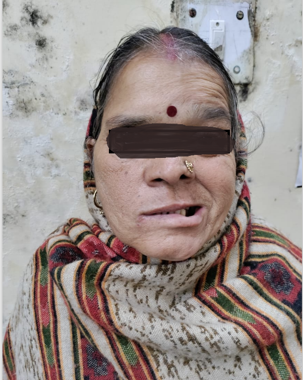
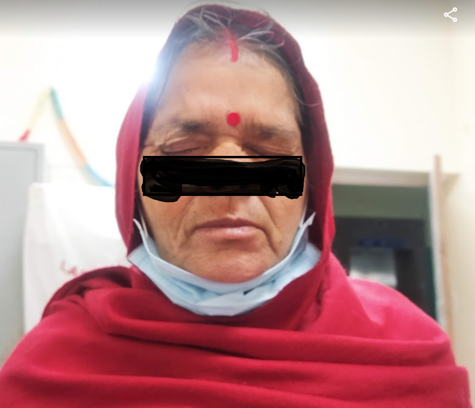
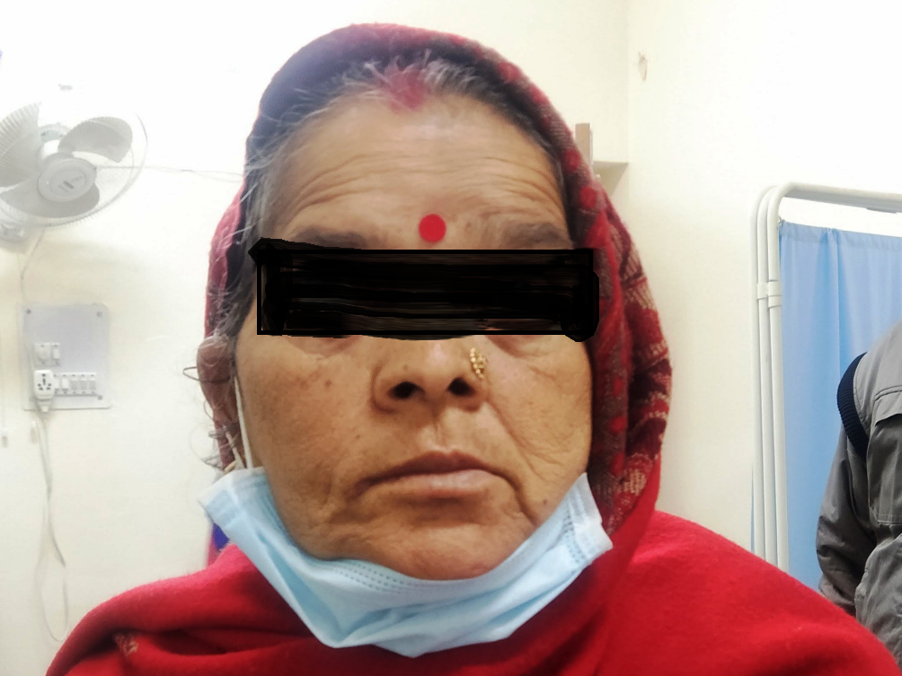
A 60 years old female patient presented in ENT OPD with a history of pain, swelling Rt side of the face for 3 days and inability to close her right eye for 2 days. She was a known case of diabetes mellitus and hypertension. She was taking treatment for her ailments but diabetes was uncontrolled with her HbA1C being 10.9%. Her TLC was 9.57x10³ with neutrophilic dominance. Rest of her routine blood investigations were in normal range. Her clinical examination revealed right side complete facial nerve palsy (Figure 1) with tenderness and small, diffuse swellings in right parotid and submandibular glands areas. The rest of ENT examinations was normal. Her ultrasound showed increased vascularity in right parotid and submandibular glands and both were bulky as compared to left side. As the patient was a known case of uncontrolled diabetes mellitus, she was admitted in the ENT ward for management of right sided facial palsy with acute parotitis and submandibular adenitis. She was put on IV, third generation Cephalosporins along with aminoglycosides and anti-inflammatory drugs. She was put on oral corticosteroids and methylcobalamins after consultations with medical specialists about her diabetes and hypertension status. Patient showed recovery from 2nd day onwards so no further radiological investigations were done in relation to her facial nerve status. After 5 days her right sided facial swelling and tenderness started subsiding and facial nerve function was also recovering. She was discharged on tenth day with no facial mass/tenderness, blood sugar was controlled and wrinkling started on right sided forehead (Figure 2), with advice to continue physiotherapy. Her corticosteroids were tapered in next twenty days and then further low dose oral corticosteroids and nervitonic for next 20 days. Her facial nerve function completely recovered after that (Figure 3).
Discussion
As a rule the combination of parotid gland lesion and facial nerve palsy is a sign of malignancy, and the clinicians should be aware that, on rare occasions, facial nerve dysfunction may result from benign parotid gland disease. In some situations, inflammation surrounding a benign neoplasm may account for the facial nerve paralysis. Marioni G et al.5 have observed that suppuration and abscess formation in the parotid are also benign extremely rare causes of facial nerve palsy and very few cases have been reported.
The etiology of paralysis remains unknown, although Moore GF6 has proposed that facial nerve dysfunction may develop due to a virulent microorganism, perineuritis or nerve compression. Streppel et al.7 reported a case of facial nerve palsy due to an infected epidermoid cyst which did not enclose the facial nerve. They proposed that the inflammation spread into the fallopian canal through the stylomastoid foramen causing a metabolic imbalance similar to the supposed vicious circle for Bell’s palsy.
As per Hajiioannou JK et al.8 acute suppurative parotitis can be attributed to salivary stasis due to block in the Stensen’s duct or a decrease in the production of saliva, poor oral hygiene, lack of mastication. They further observed that acute suppurative parotitis is usually seen in patients with diabetes.
Similarly, Brook I9 has concluded that of all the salivary glands, the parotid gland is most commonly affected by bacterial parotitis, which is generally caused by Staphylococcus aureus, Strepto (rarely), and anaerobic bacteria including Peptostreptococcus, Bacteroid species. Risk factors include age, dehydration, intubation, poor oral hygiene, anticholinergic and other drugs that reduce salivary flow, malnutrition, oral cavity neoplasms.
Pattanshetti VM et al.10 has advised that imaging of the gland should be indicated in patients who do not respond to medical therapy within 48–72 hours. They further suggested that ltrasound scan (US) is the initial imaging modality of choice for the assessment of palpable abnormalities of the parotid gland or suspected parotid calculus disease, as it has the ability to distinguish a cystic from a solid lesion, a diffuse parotid swelling from lymph nodes or an abscess and detect malignant features of the examined lesions. In acute inflammatory diseases sonography can differentiate between obstructive or non-obstructive sialadenitis as well. However ultrasound has limited visualization of the deep lobe of parotid gland which is obscured by the mandible. In our case the patient initial ultrasound scan was done and as she improved no further imaging was planned.
According to Kristensen RN et al.11 parotitis complicated by facial nerve palsy should be treated with empirical antibiotics and by a conservative approach such as hot compresses, analgesics, sialogogues, and hydration. Similarly, we started with higher antibiotics and other adjunctive therapy and patients improved.
Kanzhuly MK et al.12 have described that mortality rate in suppurative parotitis is 20% - 40%. They proposed early surgical intervention in parotid infections when there was large/increasing pus collection as demonstrated by USG/Imaging or there was a lack of improvement in clinical status. In our case, the early conservative intervention controlled the infection and prevented further complications.
Conclusion
Facial nerve paralysis accompanying benign parotid lesions or acute infections is a rare condition and malignancy must be ruled out with proper investigations while starting the treatment. Ultrasonography is the initial modality of choice for evaluating palpable abnormalities of the parotid gland, as it cheap, easily available. A complete recovery of the facial nerve paralysis the commonest outcome, and duration of symptoms may vary as already discussed.


