Introduction
The oral mucosa is constantly under the influence of various internal and external stimuli so it exhibits a range of developmental disorders, irritation, inflammation and neoplastic conditions.1
The oral cavity is the most common site for various tumours and tumour like lesions.2 Tumours may be benign or malignant. Various studies have been done for malignant tumours whereas very few studies have been done on benign tumours and tumour like lesions of oral cavity. 3
Benign tumours includes fibroma, lipoma, hemangioma, pleomorphic adenoma and leiomyoma. Tumour like lesions include pyogenic granuloma, mucocele and epulis. Oral lesions are encountered by ENT surgeons, dentists, dermatologists. Hence knowledge of benign tumours and tumour like lesions in terms of incidence, presentation and histopathological features are beneficial for better diagnosis and treatment.4
Material and Methods
A descriptive cross sectional study was carried in the department of otorhinolaryngology and head and neck surgery, Sri Siddhartha Medical College Hospital, Tumkur from September 2018 to August 2019. The aim of the study was to know the various charactaristics of benign tumour and tumour like lesions of oral cavity in terms of site, sex distribution, appearance and histopathological features. Institutional Ethical Committee approval was obtained.
All the patients of both sex between the age group of 5 to 70 years with oral cavity tumours and tumour like lesions was enrolled in the study. Patients having Premalignant and malignant tumours of oral cavity, bony tumours of mandible and maxilla were excluded. Purposive sampling technique was applied and sample size was 93.
Patients included in the study were thoroughly explained regarding nature and purpose of study and informed written consent was obtained.
Detailed history pertaining to the onset, duration and progression of the lesion and its associated symptoms, habits and occupation were collected using a questionniare.
Detailed examination regarding location, size, shape, colour and consistency was done and photographs were taken to support the recorded data. Radiological investigations like CT scan and MRI scan was done whenever necessary.
Wide excision was done in major OT after routine pre-op investigations and excised specimen was sent for histopathological examination.
Observation and Results
Tumour like lesions
48 cases of tumour like lesions was seen and confirmed by histopathological examination after intra-oral wide excision under local anaesthesia in major OT.
Table 1
Frequency of tumour like lesion
|
Tumour like lesion |
Cases |
|
Pyogenic granuloma |
26 |
|
Mucous retention cyst |
18 |
|
Fibrous epulis |
4 |
26 cases were pyogenic granuloma. Patients presented with symptoms like swelling and difficulty in speech. Lower lip was the most common location followed by gingiva and buccal mucosa. Males were more encountered than females. On examination they appeared as reddish smooth mass with ulcerated surface and non tender on palpation.
Mucous retention cyst was found in 18 cases. Females were encountered more than males. Buccal mucosa was the commonest site followed by mucosal surface of lower lip and floor of mouth. Most common complaint was excessive salivation. On examination they appeared as pinkish nodular swelling with smooth surface.
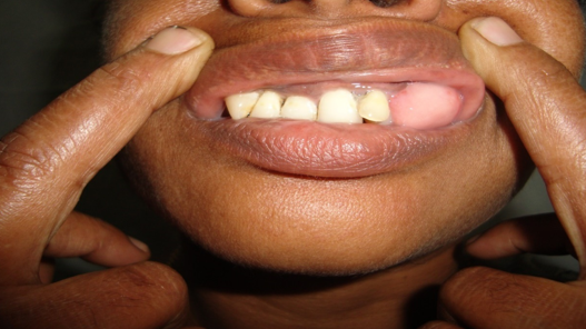
Fibrous epulis was seen in 4 cases.3 cases were seen in female patients and all cases were seen on gingiva. History of denture usage was present in all cases and on gross examination all cases presented as painless fleshy nodules.
Benign tumours
45 Cases of benign tumours of oral cavity were seen
Table 2
Frequency of Benign tumours
|
Benign tumours |
Cases |
|
Lipoma |
16 |
|
Hemangioma |
12 |
|
Pleomorphic adenoma |
9 |
|
Irritational fibroma |
6 |
|
Leiomyoma |
2 |
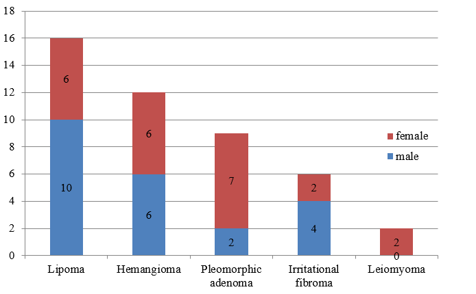
Lipoma was the most common benign tumour present in 16 cases. Buccal mucosa was the common site followed by tongue, gingiva and labial mucosa. Males were more encountered than females. It presented as yellowish sessile mass with smooth surface and soft on palpation. Size varied from small to very large swelling. Small lesions were excised under local anaesthesia whereas general anaesthesia was used for large cases.
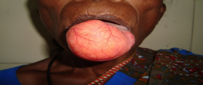
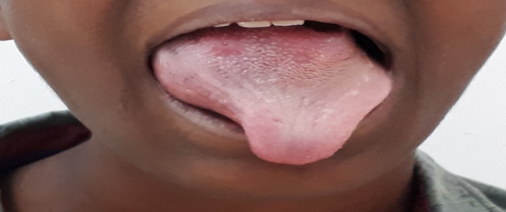
12 cases of hemangioma were seen. Tongue was the most common site followed by lower lip, buccal mucosa and alveolar ridge. Males and females were equally encountered in our study. On examination they appeared as dark red to purple, soft and sessile mass with smooth surface.4 cases were seen in children. MRI was done for large lesions on tongue. Small lesions were treated by wide excision under local anaesthesia and large sized were treated using General anaesthesia. One case of large hemangioma of base of tongue was treated by sclerotherapy.
Pleomorphic adenoma of minor salivary glands were seen in 9 cases. Palate was the most common site followed by upper lip and buccal mucosa. Females were more encountered and they appeared as asymptomatic firm swellings with smooth surface. Size of the swelling was bigger compared to other benign tumours. Wide excision was done under general anaesthesia
Irritational Fibroma was seen in 6 cases. Buccal mucosa was the most common site followed by palate, tongue and gingiva. They presented as painless pink nodule and females were more encountered. Wide excision was done under local anaesthesia.
Leiomyoma was seen in 2 cases. Both cases were seen on tongue and both were female patients. They presented as firm painless mass with smooth surface. Wide excision was done under local anaesthesia
Discussion
The term oral cavity includes lips, buccal mucosa, teeth, gums, oral tongue, floor of mouth and hard palate. Since oral mucosa is constantly under the influence of various internal and external stimuli it exhibits a range of developmental disorders, irritation, inflammation and neoplastic conditions.5 Presence of teeth and odontogenic tissue and mastication makes it susceptible for trauma.1
In 1971 WHO classified oral lesions into malignant tumors, benign tumor and tumor like lesion. According to WHO 2005 classification lesions of oral cavity are classified into malignant tumors and its variant epithelial precursor lesions and benign tumors excluding tumor like lesion. 2
Benign tumours and tumour like lesions are frequently encountered in ENT practice.
In the present study 48 cases of tumour like lesion were seen.Tumour like lesions are also called reactive lesions as they are a response to a low-grade irritation or injury such as chewing, food impaction, calculus and iatrogenic injuries such as overhanging dental restorations and extended flanges of denture. 6
Pyogenic granuloma was the most common tumour like lesion seen in 26 cases which is similar to studies of smita et al, yasmin et al and hamideh et al. Males were more encountered in our study similar to prashant et al. Lower lip was the most common site in our study whereas gingiva is most affected according to smita et al, yasmin et al and hamideh et al.7, 8, 9
It was first described in 1897 by Poncet and Dor. They called it as Botrymycosis hominis. The name pyogenic granuloma was suggested by Crocker in 1903. 1
Pyogenic granuloma appears as a smooth or lobulated asymptomatic mass that is usually pedunculated or sessile. The surface is characteristically ulcerated and friable which may be covered by a yellow fibrinous membrane. Depending on the duration of the lesion its color ranges from shiny red to pink to purple with soft to firm in palpation. Most frequently affected site is the gingivae followed by the lips, tongue and buccal mucosa. 6 Histologically all PG showed numerous small and large vascular channels which are engorged with RBCs and lined by flat or plump endothelium. 1
Second most common tumour like lesion was mucous retention cyst seen in 18 cases and females were more encountered in our study similar to smita et al but differs in the most common affected site. Buccal mucosa in our study and lower lip in their study. Mucoceles are mucous cysts related to obstruction or trauma of the minor salivary glands. They typically appear as dome shaped mucosal swellings that range from 1 to 2 mm to several centimeters in size. It is often seen in young individuals and lower lip is the classic location.
Microscopically they were classified as Extravasation type & Retention type. Extravasation type showed foci of stromal reaction due to spillage of mucous from a traumatically injured minor salivary gland whereas retention cyst shows mucous filled cyst completely lined by cylindrical, cuboidal or flattened cells. 1
4 cases of epulis were seen in our study. In our study 3 cases were encountered in females and most common site was gingiva which is similar smita et al, Maryam et al and altaf et al. Epulis fissuratum or denture-induced hyperplasia is a reactive lesion of the oral cavity caused by low grade chronic trauma from ill-fit denture. The lesion appears as an asymptomatic single fold or multiple folds of hyperplastic tissues in the alveolar vestibule along denture flanges with a smooth surface and soft to firm consistency. It is more prevalent in the maxilla than the mandible especially on the facial aspect of the alveolar ridges. Two-thirds to three-fourths of all cases have been found in females. 6 Microscopically it appears as mass covered by stratified squamous epithelium and composed of proliferating fibroblasts arranged haphazardly and at places in short bundles. 1
45 Cases of benign tumours were seen in our study
Lipoma was the most common benign tumour in our study seen in16 cases. Males were more encountered and most of the patients are over 40 years similar to Allon et al but differs to their study in the most common affected site. Buccal mucosa in their study and tongue in our study. It presents as a slow growing nodular, sessile or even pedunculated mass with a smooth surface and fluctuant to soft in palpation. The superficial lesions usually show yellowish hue while more deeply seated ones appear as a pink mass. Lipoma varies in size from small to large masses. The buccal mucosa and buccal vestibule are the most commonly affected sites followed by tongue, floor of the mouth, and lips.6, 7
12 cases of hemangioma were seen in our study with equal sex distribution. Females were encountered more in study by manisha et al. Tongue was most affected site in our study similar to Allon et al.
Hemangioma are most common in the lips, gingivae, tongue, and buccal mucosa. Clinically, it is characterized by an asymptomatic, soft, smooth or lobulated sessile mass with various sizes from a few millimeters to several centimeter. The color of the lesions ranges from pink to red purple and tumor blanches on the application of pressure. There is a tendency for female predilection. Treatment of hemangioma depends on its size and location. Small lesions are treated by sclerotherapy, conventional surgical excision, laser therapy and cryotherapy. In large cases, treatment should include embolization or obliteration of the lesion and the adjacent vessels. 6
9 cases of pleomorphic adenoma were seen in our study. Females were more encountered and palate was the most common site in our study similar to Allon et al. Pleomorphic adenoma or mixed tumor is a benign salivary gland tumor. Corresponding to minor salivary glands the palate is the most affected site followed by lips, buccal mucosa, tongue, floor of the mouth, pharynx, retromolar area and the nasal cavity. It appears a unilateral, asymptomatic, slow growing, firm, nodular or dome-shaped mass covered by a normal-colored mucosa. The surface of the lesion is usually intact. The size of the lesion varies from 1 to 5 cm. In large cases bone resorption has been reported. Majority of patients have been reported in their fifth to sixth decades of life. Lip lesions tend to occur at an earlier age than that of other locations.6
6 cases of irritational fibroma were seen in our study. Females were more encountered and buccal mucosa was the most common site similar to Maryam et al and altaf et al. It appears as an asymptomatic, pedunculated or sessile exophytic lesion with a smooth surface. It can be firm and resilient or soft with spongy consistency. The most commonly affected site is the buccal mucosa along the line of occlusion followed by labial mucosa, tongue and gingivae. There is a female predilection with the majority of cases being reported in the fourth to sixth decades of life. 6, 10
2 cases of leiomyoma were seen in our study. Both cases were seen on tongue and both were female patients similar to Allon et al. It is a benign neoplasm of smooth muscle. It usually presents as firm sessile painless mass with smooth surface. Most common location is tongue and lips. Microscopically it appears as encapsulated mass with cellular proliferation of smooth muscle cells.


