- Visibility 100 Views
- Downloads 17 Downloads
- DOI 10.18231/j.ijoas.2021.029
-
CrossMark

Haemangioma Pinna- A rare case report
Introduction
Haemangioma is a benign vascular soft tissue tumors found frequently in the head and neck region. [1] Haemangioma is a blood vessel tumour and it comprises of two greek words – haema means blood, angio means vessel & the suffix oma means tumour. It is a tumour of infancy & most cases appear during the first days or weeks of life & resolve within the age of 10 years.[2] Vascular anomalies of the head and neck didn’t have consistent nomenclature in past and the term “haemangioma” has often been used to describe any vascular lesion and is commonly preceded by descriptive but unhelpful terms, such as “cavernous” and “capillary”. In 1982, Mulliken and Glowacki developed a new system of classification of cervicofacial vascular anomalies that correlated clinical presentation and growth behaviour with histolopathological features. Vascular anomalies were thus divided into two main categories: haemangiomas and vascular malformations[3] Haemangioma in the pinna is a rare condition. Haemangioma in the pinna usually is not associated with complications, until cartilage in it may break down on the surface. [4] We are presenting a case of 61 years old female who presented with a history of non-compressible/cystic, nonpulsatile mass with minimal discoloration on right pinna for few years and only on HPE, the diagnosis of Capillary haemangioma was made.
Case Report
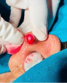
A 61 years old lady came to ENT OPD at our Institute with a small mass around 1.5 cm in diameter, on right pinna for few years. It was cystic and non-pulsatile mass which was not tender. It was very slow growing and she came for its removal only for cosmetic purpose. It was having small area of discoloration which was non blanching. There was history of ear piercing in younger age, but no recent history. Provisional diagnosis of lipoma/sebaceous cyst (though un common here)/foreign body granuloma was made which was to be confirmed by excision biopsy. After all relevant investigations for excision under local anesthesia, patient was taken for surgery. On exploration, the mass bright red tumorous tissue and not like any cyst ([Figure 1]). Mass was not attached to auricular cartilage. It was bleeding heavily. Bleeding was controlled with pressure and chemical cautery. Haemostasis secured and wound closed ([Figure 2]). Specimen was sent for histopathological examination. Microscopic examination revealed nodular fibro collagenous tissue with capillaries and small sized blood vessels with hyalinised vessel wall. No atypia was seen and a diagnosis of haemangioma was made. Patient was followed up later on and there was no recurrence or any complication.

Discussion
Vascular tumoral lesions of pinna are uncommon findings in ENT practice. Meher R et al have described haemangioma as a vascular lesion usually found in the dermis and further said that haemangiomas are neoplastic, and affect females more than males.[5] Richter GT et al, in their study on haemangiomas and vascular malformations have found that haemangioma are vascular tumours, which grows by cellular hyperplasia. [6]
A pyogenic granuloma is an acquired benign tumor, principally of the skin and mucosa; it is also termed a capillary haemangioma by reference to the pathological classification. The tumor etiology remains uncertain but may involve frequent ear-picking and changes in hormone levels.[7]
Rachappa MM et al, have written in their study that haemangiomas occur in skin of 4-10% of Caucasian new born with 3- to 5-fold greater incidence in females and also admitted that differentiation between a capillary haemangioma and pyogenic granuloma is somewhat unclear at this time. [8] Mills SE et al, suggested that the scientifically accurate term for pyogenic granuloma is the lobular capillary haemangioma.[9]
Rabbani SMG et al, have quoted Haggstrom AN et al [10] in their paper to say that haemangiomas are tumours of infancy and childhood, most of them appear during the first days or weeks of life and resolve within the age of 10 years. They further described that haemangiomas which do not resolve may present as malformations of endothelial lining cells of blood vessels. If such haemangiomas are just under skin they may present as a bluish swelling even it may present as a flat red or pink area. [4]
After going through all the above, we came to the conclusion that in this case, the lesion which was a haemangioma on histopathological examination, is a lobular capillary haemangioma/ pyogenic granuloma. It is not found in literature that any of such lesions occurs at pinna. Most of the cases found in literature were in the external auditory canal and the etiology for such lesions was proposed by Song K et al [7] as due to repeated trauma by ear picking. Hence they are acquired lesions and not congenital ones which according to Haggstrom AN et al [10] appear within few days of life.
Similarly in our case the presentation was at the age of 61 years which goes against theory of congenital lesion. Further there was history of ear pricking in the past and histopathologic findings of haemangioma. Hence it was concluded that it was a case of acquired lesion which was a haemangioma in accordance with findings of Song K et al. [7] Such small lesions in accessible areas have to be dealt with simple excision only. Further the findings on histopathological examination confirms the age old principle that any excisional biopsy must be sent for histopathologic characterization.
Conflicts of Interest
The authors declare that there are no conflicts of interest regarding the publication of this paper.
Source of Funding
None.
References
- F Martines, D Bentivegna, E Maira, S Marasa, S Ferrara. Cavernous haemangioma of the external auditory canal: clinical case and review of the literature. Acta Otorhinolaryngol Ital 2012. [Google Scholar]
- A N Haggstrom, B A Drolet, E Baselga. Prospective study of infantile haemangiomas: clinical characteristics predicting complications and treatment. Pediatrics 2006. [Google Scholar]
- J B Mulliken, J Glowacki. Haemangiomas and vascular malformations in infants and children: a classifi cation based on endothelial characteristics. Plast Reconstr Surg 1982. [Google Scholar]
- Smg Rabbani, M A Chowdhury, N Yasmeen, M Malakar. Haemangioma left pinna with extensive,multiple skin ulcerations. Bangladesh J Otorhinolaryngol 2010. [Google Scholar]
- R Meher, S Varshney, H C Pant. Arteriovenous malformation related to the pinna. Hong Kong Med J 2008. [Google Scholar]
- G T Richter, A B Friedman. Haemangiomas and Vascular Malformations: Current Theory and Management. Int J Pediatr 2012. [Google Scholar] [Crossref]
- K Song, J Lee, M J Park, H Y Lee. A Case of Bilateral External Auditory Canal Pyogenic Granuloma in a Pregnant Woman. J Audiol Otol 2018. [Google Scholar] [Crossref]
- M M Rachappa, M N Triveni. Capillary haemangioma or pyogenic granuloma: A diagnostic dilemma. Contemp Clin Dent 2010. [Google Scholar]
- S E Mills, P H Cooper, R E Fechner. Lobular capillary haemangioma: the underlying lesion of pyogenic granuloma. A study of 73 cases from the oral and nasal mucous membranes. Am J Surg Pathol 1980. [Google Scholar]
- A N Haggstrom, B A Drolet, E Baselga. Prospective study of infantile haemangiomas: clinical characteristics predicting complications and treatment. Pediatrics 2006. [Google Scholar]